Saccoglossus microinjection notes
Injection method: Rindy Jaffe injection manual
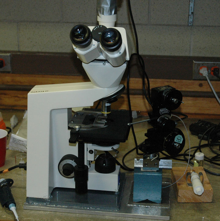
Injections being done with a 10x 0.3 NA objective on an upright
microscope with a
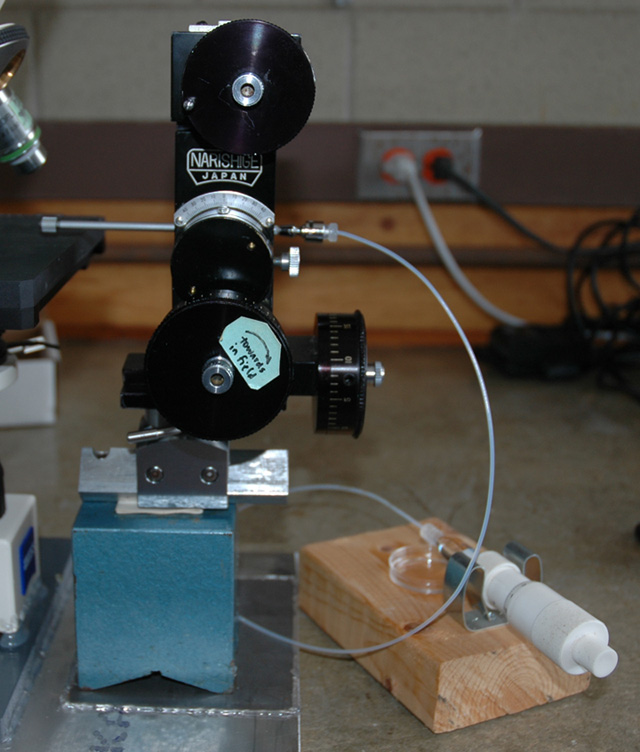
manipulator that moves in xyz directions relative
to the microscope field (Narishige SM-20). The pressure was applied by
a screw controlled hydraulic syringe (Gilmont)
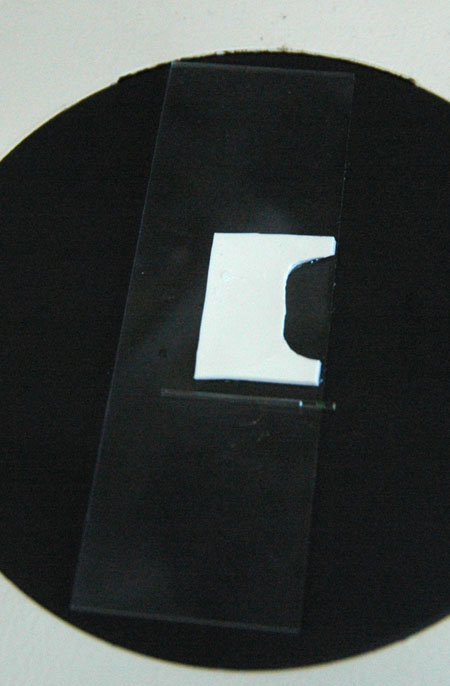
Injection chamber, made of silicon rubber (~750 µm
thick). Note also the loading capillary with greenish siRNA.
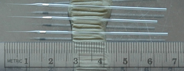
Needles used for injection note drop of mercury in
the needle, this is backfilled. The tip is broken slightly in the microscope
(1-2 µm approximately) and the mercury is pushed to the front.
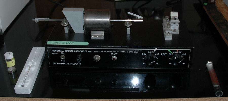
Needles were pulled on an Industrial Sciences puller
Some
preparation:
--Several sperm packets in 10-20 ml sea water in a glass
dish. These can last 1-1.5 hrs but often less. Sperm may last longer if sperm
packet remnants are cleared out of the sperm dish.
--Preparation of solutions to inject. Typically a 5 ul aliquot of siRNA. To
this, add 0.5 ul of 0.5 mg/ml calcein. Store in freezer. Make loading capillary
with
0.3 ul siRNA.
--Agarose dishes. Make 1% agarose (electrophoresis grade) in water. Melt in
microwave, then pour into 35mm petri dishes to just cover bottom and let solidify.
Add filtered seawater for 5 min to equilibrate salts. Replace with fresh
seawater.
To inject:
--Use fresh needle for each run
--Pre-load with 14 div of siRNA (1.4 mm). Am not using oil between mercury
and siRNA. Break the tip to a minimum size, very often it is difficult to load
the
oil cap and requires a second break.
--Pipet 100-400 ul sperm into a 35 mm dish with sea water
(start with 100 ul, increase the amount as time goes on)
--Add 5-8 eggs to the 35 mm dish and swirl
--Start timer (this is about 10 sec after adding eggs and moving over to the
other table). After 15 seconds, transfer to the silicon rubber injection chamber,
tilt to get
eggs
at back,
put on a glass
cover
slip
on to. Ideally, all eggs are fertilized when first looked at. If this is not
so, then try leaving the eggs in the dish for 30 seconds before transfer to
injection chamber. Other things to try: get more sperm packets, get new eggs.
--First injected egg, at the bumpy surface stage, is usually the easiest. Second
egg
is often successful. Third egg, which is between 2-3 min after fertilization,
is
the least successful, due to lysis on re-positioning the needle in the embryo,
during injection,
or especially on withdrawal. Very slow, step-wise (2-3 steps) withdrawal may
help, but it might be unavoidable. Eggs between 3-5 min are generally successful.
After ~5 min, the cortex indents further in. Fast withdrawal seems better than
slow. Usually continued the last injection when the timer goes off at 6 min;
this usually is successful, but ones afterwards are probably much less than 50%
success rate due to lysis on pull out.
--After injection, transfer the successfully injected embryos into 35 mm dish
coated with agarose. This transfer is sometimes the trickiest step. Use a 20
ul pipetman set to 10 ul and watch everything under a dissecting scope. I found
it useful to use scintillation vial bottle caps to simulate embryos, moving
them forward when injected, turning them upside down when lysed, moving them
forward to edge of table when the embryo became stuck on the needle.Using this
information about which embryos should be saved, push the cover slip gently
back until it is safe to put the pipetman tip into the chamber without violent
disruption of the chamber and disordering of the embryo positions. Write down
the siRNA, date, time of injection and number of embryos.
Egg is approximately 360 microns, which is 24.8 nl
siRNA injections were 14 div (1.4 mm) column in a needle. This corresponds
to a volume of 230 pl
So the injections were ~1% volume
siRNA was at a concentration of 50 pg/nl
Timing: one fertilizaton run per 10 minutes. 3-5 embryos injected per run. The record is 6.
Video of injections
Complete movies are too large for this web site. What we could have shown are:
needle puncture step, different kinds of lysis, behavior of the
egg at different times after fertilization, slow pull out versus fast pull
out, etc. The two movies here show only needle pull out, which is probably
the most critical step because
this is where lysis usually occurs. Note that lysis doesn't always look like
this.
Failure: egg lyses as needle is pulled out
Success
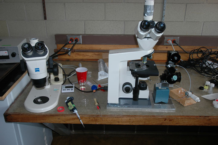
Work area around the injection microscope
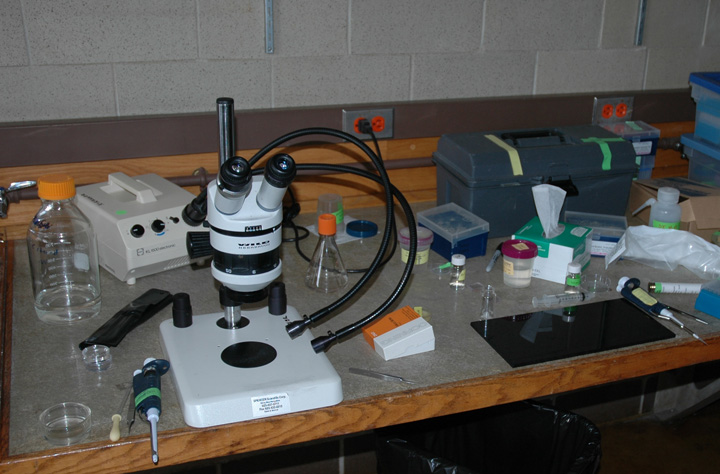
Work area for making loading capillary, sperm, etc
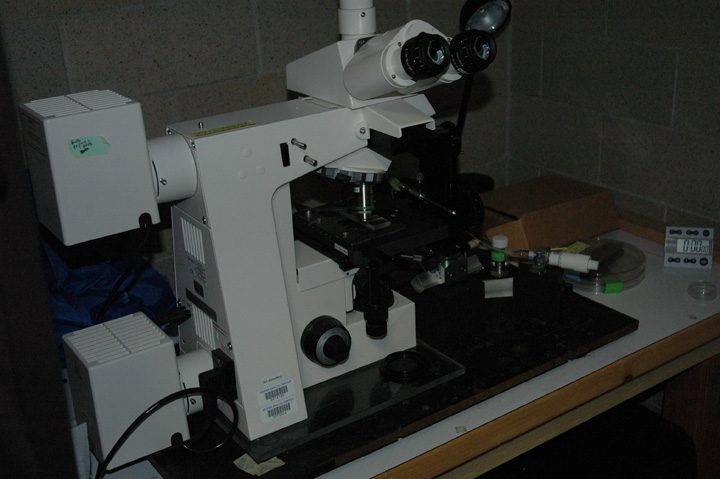
Second injection microscope, used by Linda
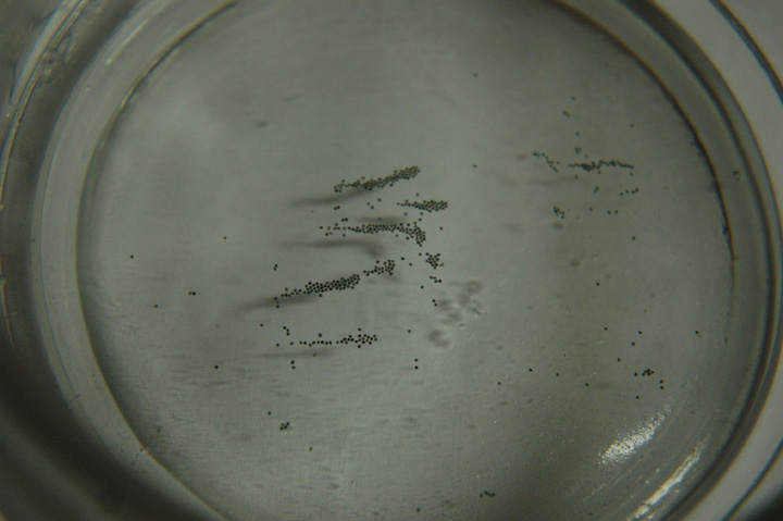
Eggs waiting to be injected
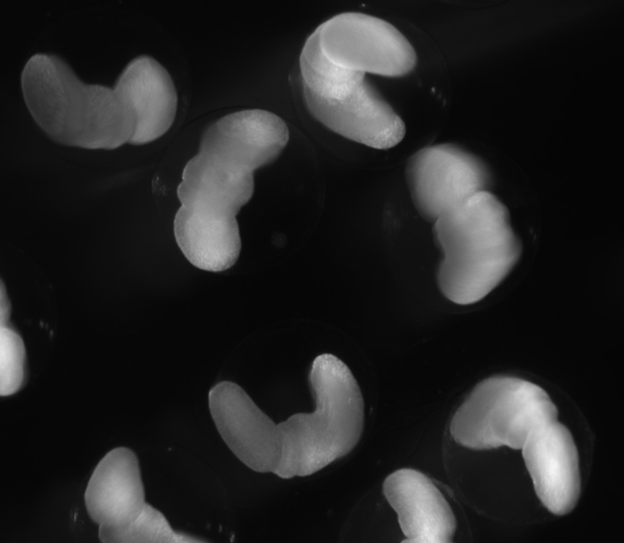
Control embryos (~ 4 days) note proboscis
BMP siRNA phenotype (~ 4 days)
Friday, September 10
--morning (11 am-12:30 pm) 13 embryos injected with siRNA for BMP 2/4 (these
all seemed to be cleaving fine around 4 pm)
--late afternoon (5:30-7 pm) 7 embryos
Saturday, September 11
--BMP (don't remember how many)
Monday, September 13
--BMP (~30 embryos)
Tuesday, September 14
--Secreted frizzled (33 embryos), wnt 8 (37 embryos)
--Wnt 3 (33 embryos)
Wednesday, September 15
--chordin (21?)
--ADMP (33)
--GFP mRNA (5)
--old BMP from last year (12)
--calcein full strength (10)
Thursday, September 16
--Otx (~30?)
--BMP (31)
--1:10 BMP (~10)
--Distal-less (~10)
--Brachyury (~10)
--ADMP / chordin mix (+30)
Friday September 17
--Chordin (~20?)
--Six-three (10)
--BMP 2.2 (32)
--1:10 dilution of BMP 2.2 (10)
--BMP 2.1 (10)
--BMP 2C (10)
Monday, September 20
--1:10 dilution BMP 2C (~16)
Tuesday, September 21
--Six-three (11)
--Brachyury (10)
Wednesday, September 22
--BMP 2C (31)
Friday, September 24
--BMP 2.2 dilutions: 1:10, 1:20, 1:40, 1:80, 1:160, 1:320 and 1:960, 3-5 of each.
--BMP 2.2 (11)
Monday, September 27
--BMP 2.2 (8); these were done at 1 hr post fertilization in DTT treated embryos.
Injections from Mon Sept 13 to Fri Sept 17 done with Linda Runft